
Have you ever noticed that tiny, pale dot resting right in the middle of your reptile’s head and wondered what hidden power it holds? To properly identify this fascinating organ, checking a bearded dragon third eye close up view reveals a small, specialized gray or translucent scale that actually functions as an ancient, light-sensing tool.
This biological marvel, scientifically known as the parietal eye, connects directly to the pineal gland, allowing your beardie to detect shadows and shifting light patterns overhead. By examining a clear bearded dragon third eye close up, any keeper can distinguish this healthy sensory dot from common issues like stuck shed, injuries, or fungal infections.
Understanding the structure of this organ helps you decode your pet’s natural defensive behaviors, especially when they suddenly freeze or bolt away as you reach into their enclosure from above. Let us dive deep into the specific physiological traits, visual features, and care methods required to master this unique aspect of reptile husbandry.
Anatomy of the Parietal Eye: A Bearded Dragon Third Eye Close Up
When you get a clean bearded dragon third eye close up perspective, the organ looks like a tiny, single opalescent scale nestled perfectly between the two primary eyes. It lacks a true iris or focusing eyelids, yet contains a primitive cornea and lens structure designed specifically to absorb ultraviolet and solar radiation.
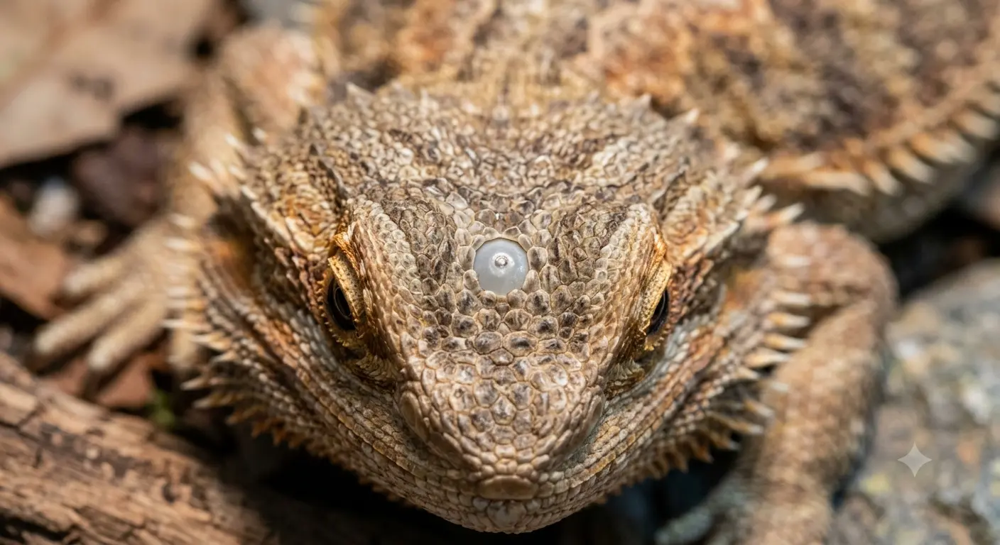
This anatomical feature does not render high-resolution images of the surrounding environment like standard vision does. Instead, it measures changes in environmental wavelengths, sending biochemical signals down to the brain to coordinate critical circadian rhythms and seasonal behaviors.
Many novice owners misinterpret this scale as a missing piece of skin or a healing wound, but close inspection proves it is an essential part of healthy anatomy. For more foundational information on how these fascinating systems interact, you can read our comprehensive analysis on bearded dragon third eye secrets to optimize your daily reptile husbandry routine.
How to Identify the Parietal Eye via Close Up Inspection
Locating this specialized scale requires a calm animal and bright, natural lighting that highlights the subtle differences in texture across the top of the skull. A proper bearded dragon third eye close up examination should reveal a tiny, smooth point that sits flush with the surrounding bumpy, spiked scales.
Healthy parietal scales are completely free of thick, crusty buildup, discolored yellow discharge, or deep deep cracks that might indicate systemic illnesses. If you notice the area looks swollen or completely obscured by layered skin, it usually indicates a localized hydration issue rather than an anatomical defect.
Keepers should never pick, scratch, or apply harsh topical creams directly to this specific zone without professional guidance. To keep your pet completely safe and well-monitored, consulting certified exotic animal experts at the Association of Reptile and Amphibian Veterinarians (ARAV) ensures you receive accurate diagnostic testing for unusual scale issues.
Distinguishing the Eye From Common Scale Conditions
It is quite common to confuse the solar-sensing dot with a stubborn patch of unpeeled skin during heavy shedding cycles. During these phases, old layers can stick to the surrounding grooves, creating a deceptive ring that mimics localized infection or abnormal discoloration.
A true bearded dragon third eye close up check shows that the natural organ retains a steady, slight shimmer even when the rest of the head looks dull and dry. If the spot looks distinctly raw, pink, or secretes any clear fluid, you are looking at a dynamic injury rather than a normal parietal scale.
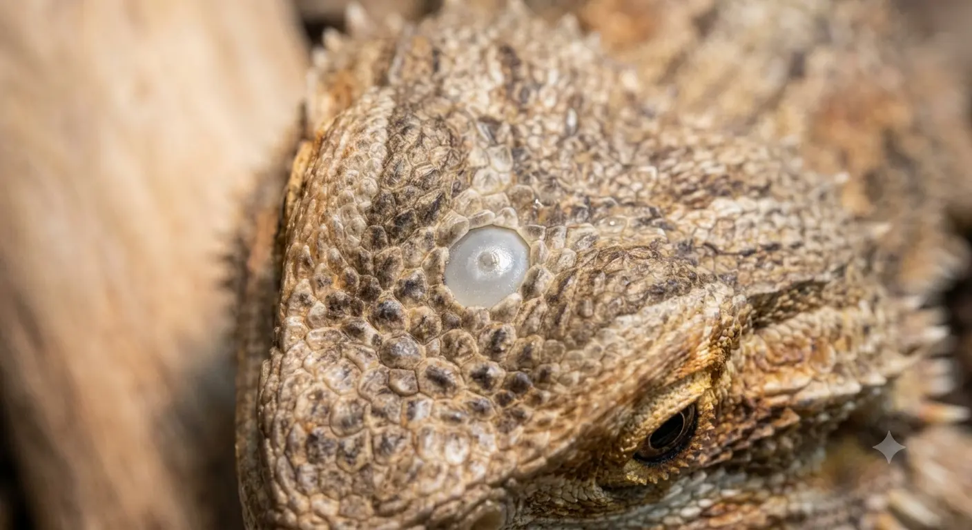
Regular photography allows you to keep an accurate visual log of your pet’s head structure over multiple months. Comparing these pictures ensures you spot slow-moving bacterial developments before they can compromise the delicate underlying neurological pathways connected to the brain.
A Master Breeder’s Tale: Overcoming a Modern Scale Scare
Two years ago, a brilliant crimson juvenile named Rocky joined my growing reptile room, bringing immense energy but also a few unexpected anxieties. One chilly Tuesday afternoon, while checking his baseline setup, I noticed a tiny, distinct gray speck right in the center of his forehead.
Panic immediately took over my thoughts as I assumed it was an early stage of destructive yellow fungus or perhaps an abrasive burn from a low-hanging heat bulb. My immediate response was to grab a damp cotton swab and scrub the tiny spot with diluted betadine solution, thinking a vigorous topical cleaning would clear it up.
Nothing changed after three days of stressful scrubbing, and my beautiful lizard began darkening his beard, showing immense frustration every time my hands approached his enclosure. I felt completely defeated, worried that a hidden infection was silently spreading across his skull while my interventions only caused further behavioral stress.
By pure chance, during a late-night phone call with an old mentor who had bred desert lizards for three decades, he laughed softly at my intense panic. He explained how this specific optical spot behaves like a natural light meter, and my aggressive scrubbing was simply irritating a perfectly healthy, light-sensitive scale.
I stopped all manual interventions immediately, adjusted his overhead lighting angles, and watched Rocky return to his relaxed, happy self within a week. That stressful week taught me the true value of patient observation over reactive, uneducated treatments when managing complex reptilian features.
Functional Comparisons: Parietal Eye vs. Normal Scales
To help you quickly differentiate this critical organ from standard tissue, reviewing specific functional parameters keeps your husbandry practices highly accurate. The following matrix outlines the core attributes you will observe during a typical health inspection:
| Physical Property | Parietal Organ Scale | Standard Cranial Scale | Stuck Retained Shed |
|---|---|---|---|
| Primary Coloration | Translucent, gray, or soft white | Matches body pattern (brown/orange) | Dull, opaque, chalky white |
| Light Sensitivity | High (detects shadows/UV changes) | None (purely protective structural tissue) | None (blocks incoming ambient light) |
| Texture Profile | Completely smooth, flat, slightly glossy | Keeled, rough, or raised at edges | Brittle, raised, dry and paper-like |
| Nerve Integration | Direct connection to pineal complex | Standard cutaneous nerve pathways | Avascular dead tissue layer |
By keeping a close eye on these specific visual markers, you can avoid unnecessary vet visits while ensuring your animal remains comfortable. Any sudden deviation from these baseline parameters warrants a careful reassessment of your environment.
Environmental Optimization for Solar-Sensing Organs
Because this specialized scale acts as a primary sensor for solar energy, your artificial terrarium lights directly impact its daily neurological performance. Providing a clear gradient of high-quality UVB light allows the organ to regulate the animal’s natural hormone synthesis smoothly.
When setting up high-output T5 bulbs, ensure they span the proper distance to prevent radiation stress on the upper skull tissues. A distorted light cycle can confuse the parietal system, leading to erratic sleeping patterns, sudden loss of appetite, or general lethargy.
Always approach the enclosure from a side angle rather than casting a sudden shadow directly over the top of the animal’s head. Minimizing these artificial predatory triggers reduces systemic stress, allowing your pet to feel completely secure within their domestic environment.
In summary, capturing a clear bearded dragon third eye close up view gives you invaluable insight into the evolutionary brilliance of your pet. By monitoring this delicate structure for changes, you guarantee a healthier, more predictable life cycle for your cherished reptile companion.